


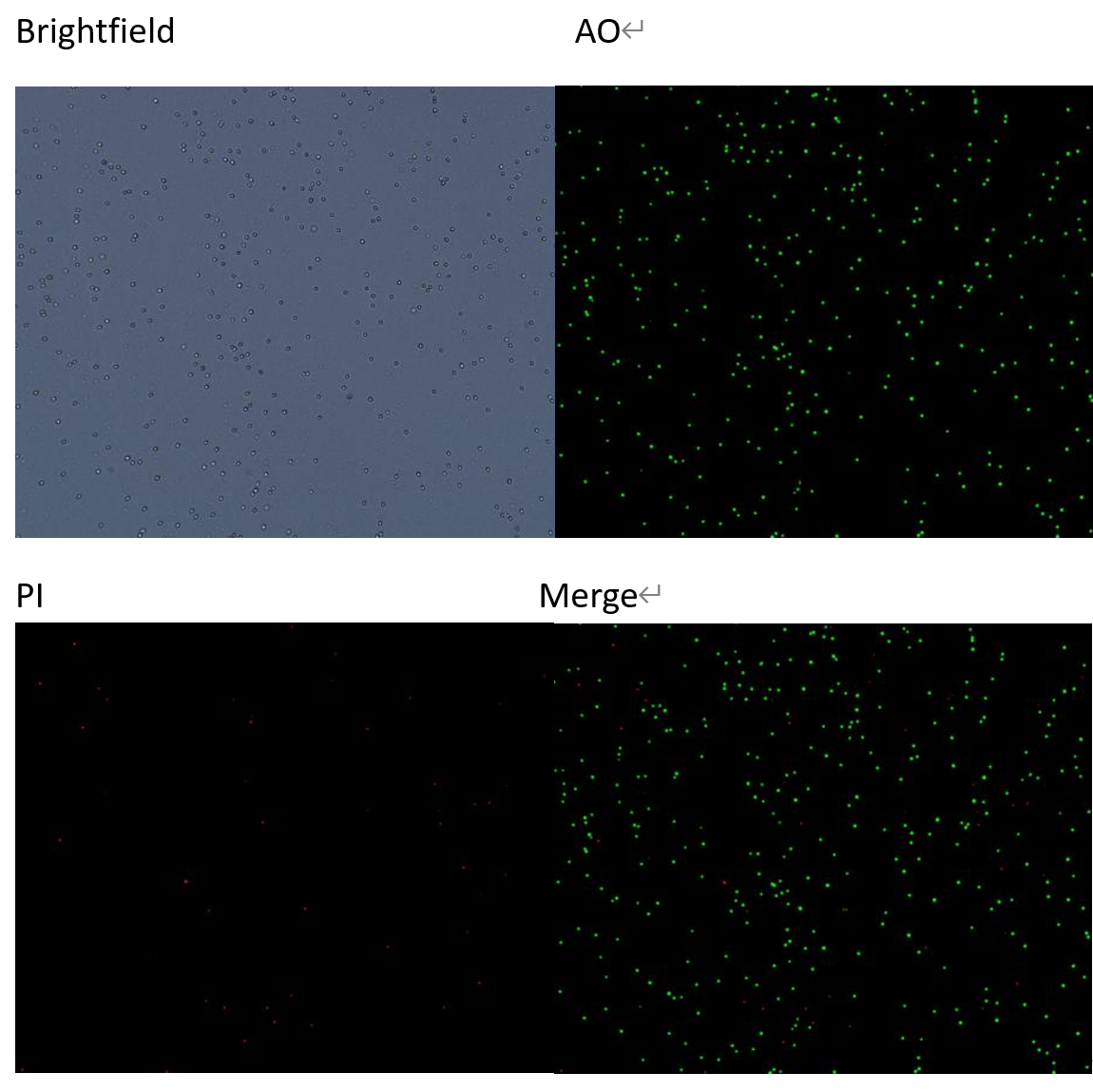
AO/PI Viability
The AO/PI dual-fluorescence staining method distinguishes live and dead cells by using two fluorescent dyes: acridine orange (AO) and propidium iodide (PI). AO can penetrate intact cell membranes and binds to the DNA and RNA of live cells, causing them to emit green fluorescence. PI can only enter dead cells with compromised membranes, where it binds to DNA and emits red fluorescence. This method helps to exclude impurities and debris from the cell samples, enabling accurate assessment of cell viability.
The fluorescence cell analyzer was validated for the stability of viability measurements after dilution of PBMC cells, and the accuracy and dilution linearity of the analyzer’s counting results within this concentration range were confirmed.
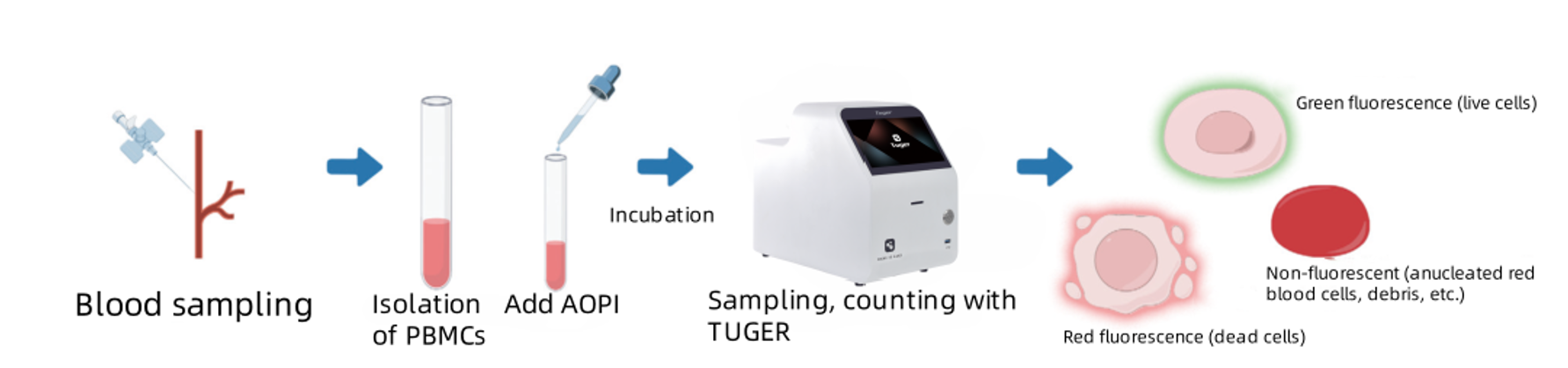
Experimental scheme:
1、After harvesting and centrifuging the PBMC cells, the cell pellet was resuspended in PBS. Manual counting determined a cell viability of 84.58%, a total cell concentration of 8.433 × 10⁶ cells/mL, and a viable cell concentration of 7.133 × 10⁶ cells/mL. The sample was labeled as Cell1;
2、Cell1 was serially diluted at 2-fold, 4-fold, 8-fold, and 16-fold to obtain cell suspensions of different concentrations, labeled as Cell2, Cell3, Cell4, and Cell5, respectively;
3、Samples at different concentrations were stained with AO/PI dyes and incubated for 1 minute;
4、The cell suspensions Cell1 to Cell5 were counted using the Tuger fully automated fluorescence cell analyzer to measure their cell concentrations and viability.
Result:
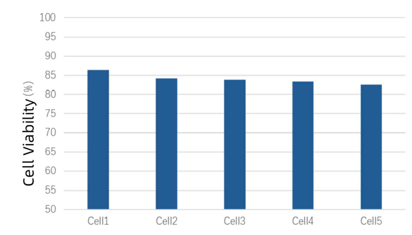
Viability of each diluted cell sample
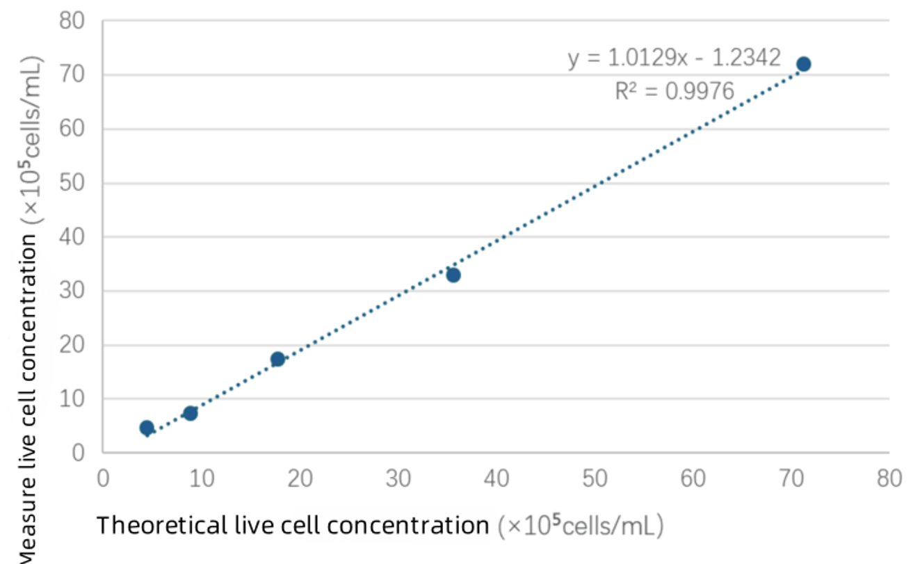
Fitting results between measured viable cell concentrations and theoretical concentrations
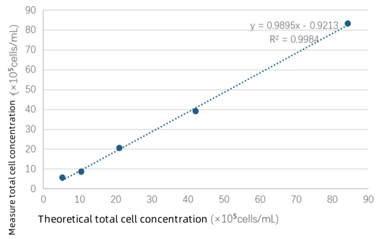
Fitting results between measured total cell concentrations and theoretical concentrations
Conclusion:
During the dilution process, the cell viability test results remained stable and were close to the values obtained by manual counting. Comparison between the theoretical total cell concentrations and measured total cell concentrations, as well as between theoretical viable cell concentrations and measured viable cell concentrations, showed that within the cell concentration range of 5 × 10⁵ cells/mL to 8 × 10⁶ cells/mL, the Newtonoptic fluorescence cell analyzer provided accurate counts with good dilution linearity. The fitting results demonstrated an R² value greater than 0.99.

National Advisory Service Hotline
Sales consultation: +86 15322248165(Whatsapp/Wechat)
E-mail:global@newtonoptic.com
R & D Center: Room 301, Floor 1, Building 1, No.38 Gaopu Road, Tianhe District, Guangzhou City, Guangdong Province

Follow Wechat Official Account
©2024 Guangzhou Newtonoptic Research Institute Co., Ltd. All rights reserved